Back Of Skull Anatomy - Human skull viewed from the back. - Stock Photo , #ad, #skull, #Human, #viewed, #Photo #AD .... Learn more about the anatomy and function of the skull in humans and other vertebrates. Inferior view of base of the skull. Functional anatomy of the skull. The skull begins to form prior to week 12 of embryogenesis. They don't move and united into a single unit.
A cartilaginous mould begins to grow this is why raising your eyebrows can create the appearance that the back of the head is moving. The skull base is the inferior portion of the neurocranium. So, the human skull consists of 23 bones. The skull or known as the cranium in the medical world is a bone structure of the head. The frontal, parietal, temporal and occipital bones are joined at the cranial sutures.
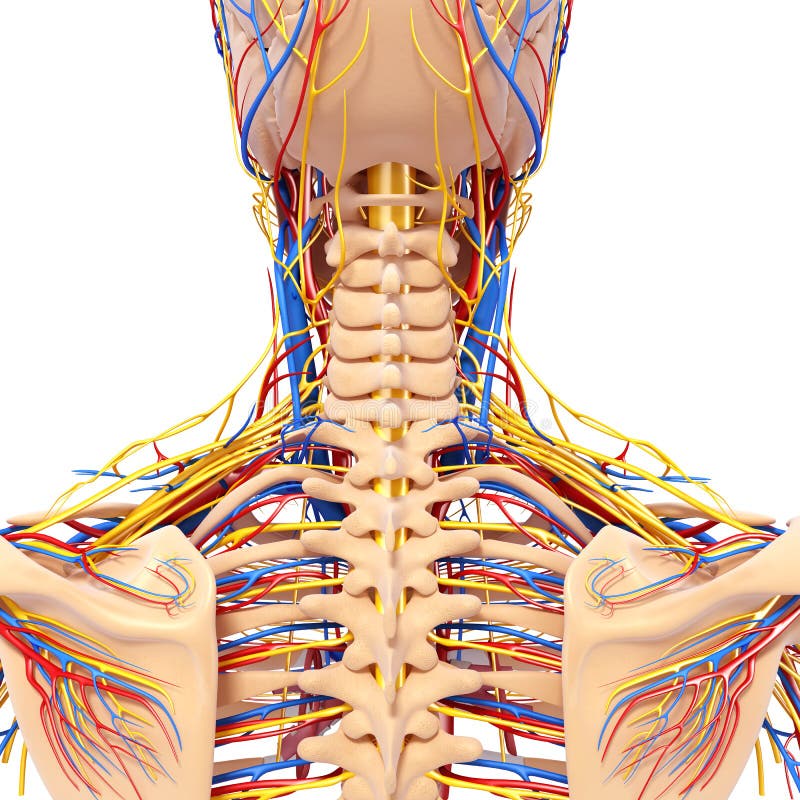
Anatomy Of Male Head Back View Circulatory System Stock Photography - Image: 26688192 from thumbs.dreamstime.com So, the human skull consists of 23 bones. The skull base is the inferior portion of the neurocranium. The bone is pierced by a large oval hole(the foramen magnum) through which runs the spinal cord. Skull, skeletal framework of the head of vertebrates, composed of bones or cartilage, which form a unit that protects the brain and some sense organs. The frontal, parietal, temporal and occipital bones are joined at the cranial sutures. Learn more about the anatomy and function of the skull in humans and other vertebrates. The anterior fossa is formed by the orbital plates of the frontal bone, cribriform plate of the ethmoid, and lesser wings of the sphenoid. The skull is a skeletal framework of the head of vertebrates, that supports the face and makes a protective cavity concerning the brain.
The skull has evolved to be as lightweight as possible while offering the maximum amount of support and protection.
Foramina inside the body of humans and other animals. Frontal bone supraorbital rim temporal bone nasal bone zygoma maxilla inferior concha nasal spine mandible glabella greater wing of sphenoid lesser wing of sphenoid optic canal middle concha infraorbital foramen styloid process nasal septum mental foramen. It offers protection to the brain, eye balls, inner ears, and nasal passages. In order to be light, the skull is made up by flat and irregular bones, and has hollow spaces called the sinuses. The skull is a unique skeletal structure in several ways: Functional anatomy of the skull. Excluding ear ossicles, it is made of 22 bones. Learn about skull base anatomy with free interactive flashcards. Bone structure differences between races. — the skull is the receptacle for the most highly developed part of the nervous system, the brain and also for the sensory organs connected with it. Human skull from the front. The anterior fossa is formed by the orbital plates of the frontal bone, cribriform plate of the ethmoid, and lesser wings of the sphenoid. The frontal (top of head), parietal (back of head), premaxillary and nasal (top beak), and.
The skull begins to form prior to week 12 of embryogenesis. The frontal, parietal, temporal and occipital bones are joined at the cranial sutures. The skull bones can be classified into two groups: Learn about skull base anatomy with free interactive flashcards. Related posts of bone of back of skull.
Back Bone With Skull Back View Stock Illustration - Image: 46887679 from thumbs.dreamstime.com Norma basalis ( anterior part , middle part and posterior part ). Human anatomy for muscle, reproductive, and skeleton. The anterior fossa is formed by the orbital plates of the frontal bone, cribriform plate of the ethmoid, and lesser wings of the sphenoid. The skull performs vital functions. The cranium (skull) is the skeletal structure of the head that supports the face and protects the brain. The axial & appendicular skeleton. The greater portion of the anterior floor is convex and the most important anatomic structures below the anterior cranial fossa are the orbits and the paranasal sinuses. Anatomy of the skull and bones of cranium on medical illustrations.
The cranium (skull) is the skeletal structure of the head that supports the face and protects the brain.
Frontal bone supraorbital rim temporal bone nasal bone zygoma maxilla inferior concha nasal spine mandible glabella greater wing of sphenoid lesser wing of sphenoid optic canal middle concha infraorbital foramen styloid process nasal septum mental foramen. Skull, skeletal framework of the head of vertebrates, composed of bones or cartilage, which form a unit that protects the brain and some sense organs. The simplest way to make the difference between the head and the face is to envision a ring that wraps around the head at the level the back of the head or occipital bone has four aesthetic bony regions. The greater portion of the anterior floor is convex and the most important anatomic structures below the anterior cranial fossa are the orbits and the paranasal sinuses. Excluding ear ossicles, it is made of 22 bones. Cranial cavity , cranial sutures. — the skull is the receptacle for the most highly developed part of the nervous system, the brain and also for the sensory organs connected with it. The skull has a single occipital condyle.7 the skull consists of five major bones: Looking at it from the inside it can be subdivided into. William is a final year medical student in australia who has taught anatomy to tertiary science and. Anatomical structures of the skull include: The bone is pierced by a large oval hole(the foramen magnum) through which runs the spinal cord. The base of the skull (or skull base) forms the floor of the cranial cavity and separates the brain from the structures of the neck and face.
Skull anatomy | with labels. The skull is the bony skeleton of the head. Learn about skull base anatomy with free interactive flashcards. Looking at it from the inside it can be subdivided into. The frontal, parietal, temporal and occipital bones are joined at the cranial sutures.
4 Ways To Relieve A Tension Headache At The Base Of Skull from www.massageaholic.com Learn skull anatomy with skull bones quizzes and diagram labeling exercises. Inferior view of base of the skull. Anatomical structures of the skull include: Looking at it from the inside it can be subdivided into. Foramina inside the body of humans and other animals. They don't move and united into a single unit. Bone structure differences between races. Human anatomy for muscle, reproductive, and skeleton.
Norma basalis ( anterior part , middle part and posterior part ).
Bone structure differences between races. Human skull from the front. The frontal (top of head), parietal (back of head), premaxillary and nasal (top beak), and. Learn about the anatomy of the skull bones and sutures as seen on ct images of the brain. The occipital bone forms the back of the skull and the base of the cranium. Learn about skull base anatomy with free interactive flashcards. The skull has a single occipital condyle.7 the skull consists of five major bones: The skull bones can be classified into two groups: It supports and protects the face and the brain. Looking at it from the inside it can be subdivided into. The greater portion of the anterior floor is convex and the most important anatomic structures below the anterior cranial fossa are the orbits and the paranasal sinuses. Cranial cavity , cranial sutures. These joints fuse together in adulthood.
Share :
Post a Comment
for "Back Of Skull Anatomy - Human skull viewed from the back. - Stock Photo , #ad, #skull, #Human, #viewed, #Photo #AD ..."


Post a Comment for "Back Of Skull Anatomy - Human skull viewed from the back. - Stock Photo , #ad, #skull, #Human, #viewed, #Photo #AD ..."